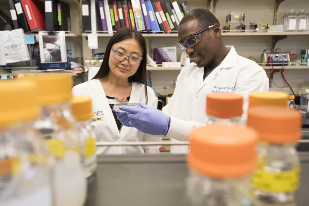
Bassnett Lab
Steven Bassnett, PhD, Professor, Ophthalmology & Visual Sciences, Cell Biology & Physiology

Research Focus: Anterior segment biology and pathology
Our research focuses on the development, biology, and pathology of tissues in the eye’s anterior segment. The work spans multiple disciplines, including biophysics, imaging, cell and molecular biology, biomedical engineering, and mathematics. Our goal is to translate the laboratory findings into improved clinical practice.
Lens Development and Growth
The mechanisms that specify lens size and shape remain largely unknown. We are currently exploring the role of physical forces, such as those exerted by the ciliary zonule, in regulating lens growth.
Glaucoma Research
We are studying exfoliation glaucoma, a type of glaucoma characterized by the buildup of protein fibrils in the eye’s drainage structures. The fibrils can block fluid flow, leading to increased pressure and retinal damage. Using mass spectrometry and high-resolution electron microscopy, we are investigating the composition and structure of the exfoliation fibrils. Our goal is to develop novel methods for diagnosing the disease, disaggregating the fibrils, and lowering intraocular pressure.
Funding
Our work is funded through grants from the National Eye Institute of the NIH. We have also received support from the Marfan Foundation, Glaucoma Research Foundation, Research to Prevent Blindness (RPB), the European Union, and the Grace Nelson Lacy Glaucoma Fund. We received NIH grants for our projects, the “Core Grant for Vision Research” and
“Structural, Mechanical, and Cell Biological Properties of the Ciliary Zonule”.
Publications

Some recent publications from the Bassnett Lab
Rodriguez, J., Tan, Q., Sikic, H., Taber, L.A., Bassnett, S. (2023). The effect of fibre remodelling on the power and optical quality of the lens. Journal of the Royal Society Interface. 20(206):20230316.
Dr Maria, A., Zientek, K.D., David, L.L., Wilmarth, P.A., Harocopos, G.J., Huang, A.J.W., Hong, A.R., Siegfried, C.J., Tsai, L.M., Sheybani, A., Bassnett, S. (2021). Compositional Analysis of extracellular aggregates in the eyes of patients with exfoliation syndrome and exfoliation glaucoma. Investigative Ophthalmology and Visual Sciences.62 (15):27.
Youkilis, J.C., Bassnett, S. (2021). Single-cell RNA-sequencing analysis of the ciliary epithelium and contiguous tissues in the mouse eye. Experimental Eye Research 213:108811.
Bassnett, S. (2021). Zinn’s zonule. Progress in Retinal and Eye Research. doi: 10.1016/j.preteyesres.2020.100902. PMCID: PMC8139560.
Shi, Y., Jones, W., Beatty, W., Tan, Q., Mecham, R.P., Reinhardt, D.P., Gibson, M.A., Reilly, M.A., Rodriguez, J., Bassnett, S. (2021) Latent-transforming growth factor beta-binding protein-2 (LTBP-2) is required for longevity but not for development of zonular fibers. Matrix Biology, 95:15-31. PMCID: PMC8276913.
Jones, W., Rodriguez, J., Bassnett, S. (2019). Targeted deletion of fibrillin-1 in the mouse eye results in ectopia lentis and other ocular phenotypes associated with Marfan syndrome. Disease Models and Mechanisms. doi:10.1242/dmm.037283. PMCID: PMC6361150.
Majtan, T., Jones, W., Krijt, J., Park, I., Kruger, W.D., Kozich, V., Bassnett, S., & Kraus, J.P. (2018) Enzyme replacement therapy ameliorates multiple symptoms of murine homocystinuria. Molecular Therapy, 26(3):834-844. PMCID: PMC5910661.
Vinberg, F., Wang, T., De Maria, A., Zhao, H., Bassnett, S., Chen, J., & Kefalov, V. (2017). The Na+/Ca2+, K+ exchanger NCKX4 is required for efficient cone-mediated vision. Elife. e24550. PMCID: PMC5515578.
Šikić, H., & Bassnett, S. (2017). The lens growth process. Progress in Retinal and Eye Research, 60:181-200. PMCID:PMC5605917.
De Maria, A., Wilmarth, P.A., David, L.L., & Bassnett, S. (2017) Proteomic analysis of the bovine and human ciliary zonule. Investigative Ophthalmology and Visual Sciences 58(1):573-585. PMCID:PMC5283081.