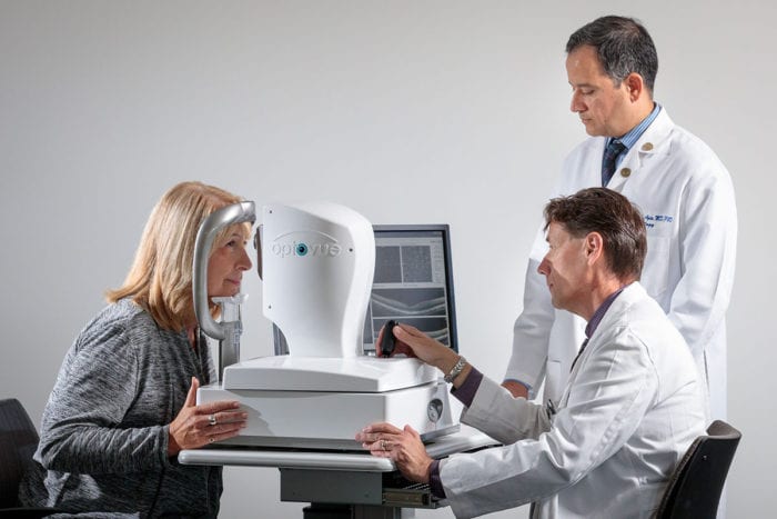

A noninvasive test may screen for disease before symptoms begin. Dr. Van Stavern is using retinal imaging as a window to the brain to develop a more accessible and affordable screening tool for Alzheimer’s disease that could become part of a routine eye exam. The retina and central nervous system are so interconnected that changes in the brain could be reflected in cells in the retina.
We know the pathology of Alzheimer’s disease begins to develop years before symptoms appear, but if we could use this eye test to notice when the pathology is beginning, it may be possible one day to start treatments sooner delaying further damage. In fact, it may be possible to one day screen people as young as their 40’s or 50’s to determine whether they are at risk for the disease. In December 2022, Dr. Van Stavern was awarded a $10.3 million grant from the National Institutes of Health to expand the effort to understand this important connection.