The Vision Function Testing Core provides Vision Function Testing (VFT) Services for mouse research including a suite of visually guided behavioral tests and in vivo electrophysiology.
In vivo electrophysiology
Electroretinograms (ERGs) measure light-evoked, electrical activity from the retina to assess rod and cone photoreceptor function, as well as signal transmission from photoreceptors to bipolar cells. Retinal pigment epithelium function can also be measured from the C-wave.
Patterned ERG uses specialized light stimulation to assess the ability of retinal ganglion cells to integrate synaptic inputs.
Visually evoked potentials (VEPs) are used to assess visual signaling between the retina and the brain. VEPS are evoked potentials recorded from the visual cortex in response to a flash of light.
Mouse visually guided behavior
Optokinetic reflex measures the ability to perform saccades in response to moving stimuli. Stimulus characteristics like spatial resolution and contrast are modified to determine visual acuity. This passive test is ideal for animals that might demonstrate broad functional deficits since it only relies on signal detection and ocular muscles.
Pupillary light response measures the ability of the pupil to constrict in response to increased light levels testing the function of a specific set of intrinsically photosensitive retinal ganglion cells. This passive test is ideal for animals that might demonstrate broad functional deficits since it only relies on signal detection and ocular muscles.
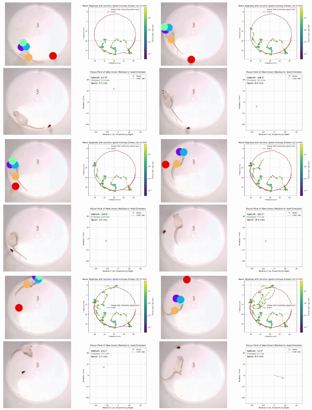
Prey capture in mice relies on high acuity binocular vision in mice. This examines the performance of spatially and functionally specialized circuits in the retina by tracking how mice capture red runner roaches.
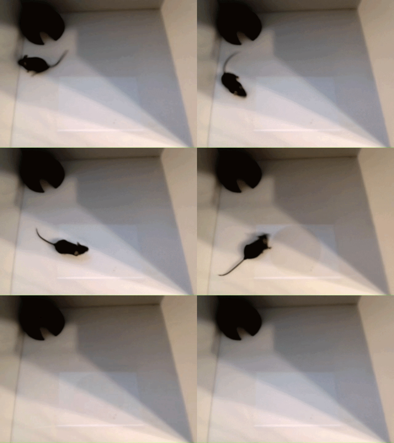
Looming response simulates how a mouse reacts to a virtual aerial predator. This test examines well-characterized retinal and brain circuits involving defined neuronal pathways.
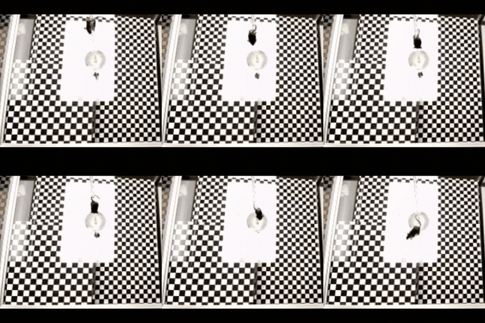
Pole-descent visual cliff measures depth perception, which requires integration of binocular signals in the brain.
Meet The Visual Function Testing Core Team
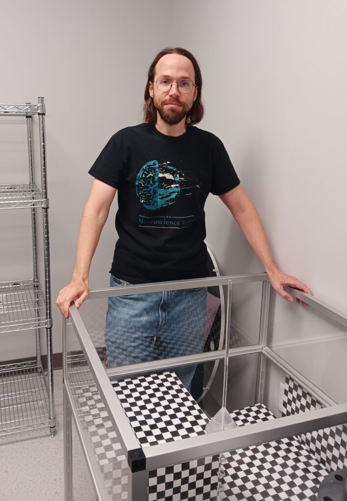
Philip Williams, PhD
Assistant Professor, Ophthalmology and Visual Sciences
Email: prwillia@wustl.edu
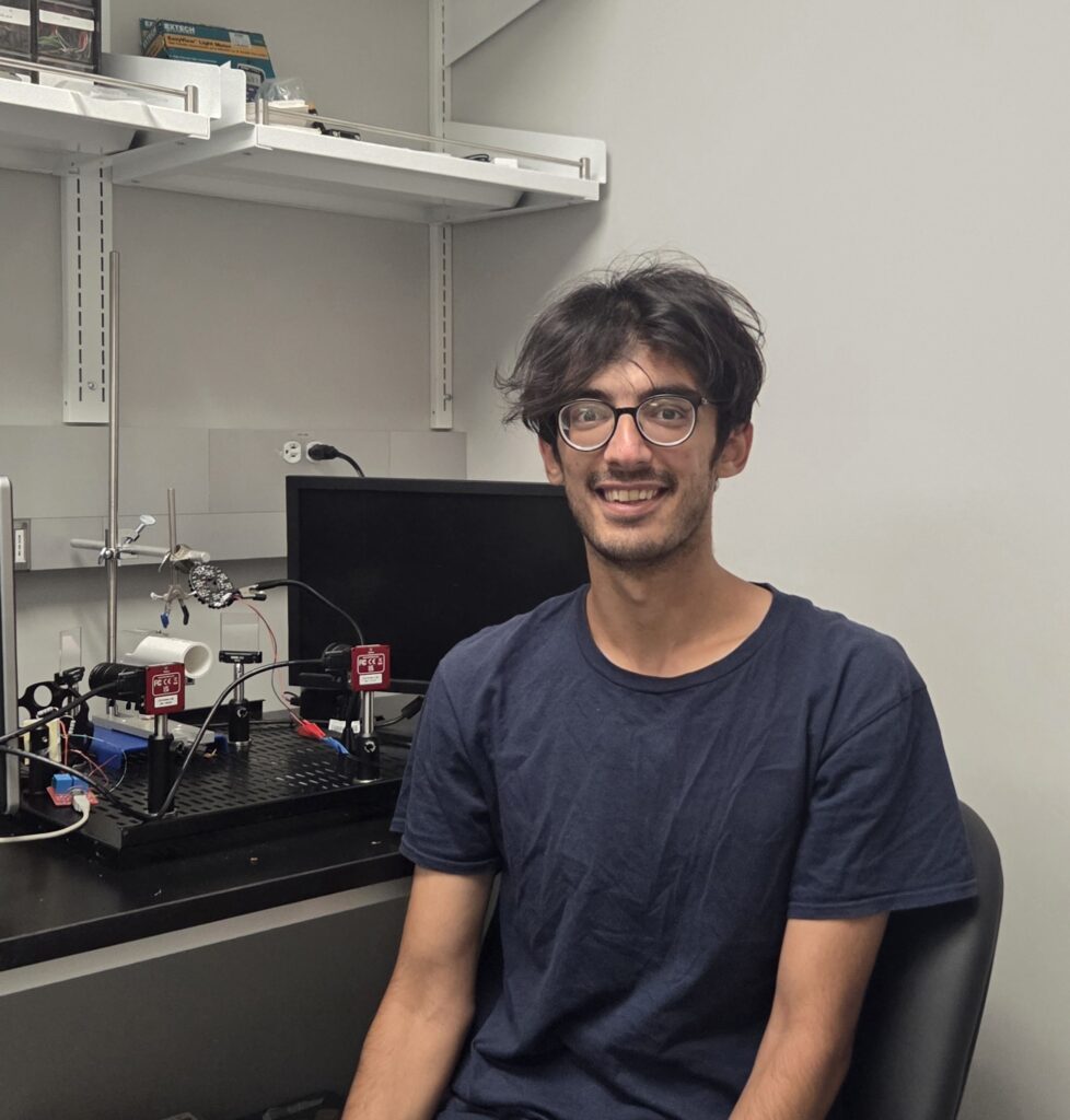
Saman Behboudi Tanourlouee, DVM
Postdoctoral Fellow, Vision Research Scientist
Email: bsaman@wustl.edu
Below is a list of major equipment available through the Vision Function Testing Core
Major Equipment | Location (MMSB) | On-line Reservation | Training required |
Celeris | 975C | Yes | Yes |
| 975D | Yes | Yes | |
Behavior Testing Center | 975A | Yes | Yes |
The department also has a Biopitgen small animal OCT system. Please see the Morphology & Imaging Core for further information.
Hours of availability:
Please contact Saman Behboudi Tanourlouee for training.
After being fully trained, you may sign-up on the FOM website (https:// www.instrumentschedule.com/fom/) to use the facility – first-come, first-serve.