A new study from the WashU Medicine John F. Hardesty, MD Department of Ophthalmology & Visual Sciences, led by Philip Williams, Ph.D., Rajendra S. Apte, MD, PhD, and graduate student Zelun Wang, offers important insight into how retinal neurons adapt their metabolism to sustain vision and respond to disease. Published in Nature Communications, the research focuses on retinal ganglion cells—critical neurons that transmit visual information from the eye to the brain—and how their energy use varies across cell types. Understanding these differences may help explain why certain neurons are more vulnerable in conditions like glaucoma and other neurodegenerative diseases and how molecular therapies that target these metabolic differences could help us make retinal neurons more resilient.
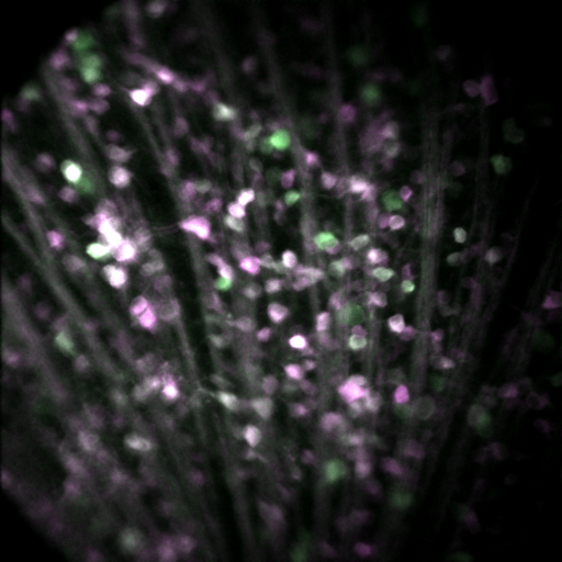
Using advanced in vivo imaging alongside high-throughput, AI-assisted analysis, the team studied these neurons within their natural environment. Their findings reveal that even closely related neurons demonstrate unique metabolic profiles, influencing how they function and survive under stress. This work highlights the importance of studying cells in context and showcases innovative approaches that combine imaging and artificial intelligence to uncover new biological insights.
“Neural tissues are highly specialized to supply nutrients to energy hungry neurons. However, little is known about how neurons themselves customize their metabolism to support their complex functions,” said Philip Williams, Ph.D. “In this study, we show that a population of similar yet diverse neurons demonstrate a range of metabolic specializations, and these characteristics influence their survival during degeneration.”
“Using novel in vivo imaging techniques with high-throughput AI-assisted analysis, we were able to investigate the metabolism of these neurons in their natural tissue microenvironment and reveal key insights into energy expenditure related to their signaling,” added Zelun Wang.